
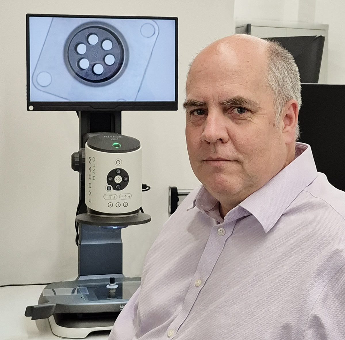

Could precision imaging evolve without eyepieces? From ergonomics, modularity, to AI-driven precision—Vision Engineering’s Steve Sanderson reveals how cutting-edge microscopy is transforming electronics and healthcare to EFY’s Akanksha Sondhi Gaur.

Q. Can you start by giving us an overview of Vision Engineering and the solutions you provide in microscopy?
A. Vision Engineering has been at the forefront of optical and digital microscopy for over six decades. Based in the UK, just outside London in Woking, we specialise in designing and manufacturing a range of stereo microscopes, optical measurement systems, and digital inspection microscopes.
All of our design and manufacturing processes, from R&D (research and development) to quality control, happen in-house. Besides, more than 90 per cent of our products are exported worldwide, for industries such as electronics, precision machining, medical devices, plastics manufacturing, forensics, and aerospace.
Q. Which industries benefit most, and what are some unique applications?
A. As I said, our microscopes support many industries. Law enforcement detects counterfeits by analysing surface markings, while medical manufacturers ensure precision in life-saving devices. High-speed sectors like semiconductor and consumer electronics rely on our solutions too. By combining optical expertise with digital advancements, we help industries transition to smarter, more efficient quality assurance processes

Q. What makes India a key market for you, and how do you compare its potential to your global customer base?
A. The increasing demand for advanced inspection tools in electronics, automotive, and medical industries makes India a key market for us. The country is at the forefront of technological advancements, with a tremendous growth opportunity. While I do not have exact figures, we see a thriving ecosystem of innovation and high-tech manufacturing.
We also have a dedicated team in India, working closely with organisations like ISRO (Indian Space Research Organisation). Our microscopes have played a role in India’s space exploration efforts, including recent lunar missions. Being part of such groundbreaking developments is both an honour and a testament to our commitment to innovation.
Q. What is the role of your company in India’s growing electronics market?
A. It is a crucial market for us, and we have distribution partners who are very able to help customers find the right solutions. Our advanced inspection technologies are being integrated into various applications, including electronics manufacturing, medical devices, and aerospace. It is so exciting to see that our technology is used in developing and quality control medical devices, ensuring they meet rigorous industry standards.
Q. In India, is your primary customer the distributor, or are you targeting end users directly?
A. While we work closely with our distribution partners, our ultimate customers are the end users—engineers, technicians, and manufacturers—who rely on our technology to enhance their work. Our goal is to make their jobs easier and more efficient.
Q. What kind of channel partnerships do you have in India?
A. We work with a strong network of distribution partners specialising in different high-precision industries, including the electronics sector.
Q. So, your products are designed and manufactured in the UK and then distributed in India through your partners. Is that correct?
A. That is right. Our core design and manufacturing operations are based in the UK, and through our distributors in India, we ensure our products reach the right customers.
Q. Do you have an R&D setup in India, or are there plans to establish one?
A. Currently, we do not have an R&D centre in India, but I must say, the passion and drive of Indian engineers and businesses to collaborate with global technology leaders are impressive. I cannot disclose specific plans, but our management has been actively exploring opportunities to deepen our engagement with the Indian market.
Q. Coming to the global, which markets are growing the fastest for you, and what is driving that growth?
A. It is the medical device sector, driven by innovations. Electronics is also evolving continuously. There are also exciting advancements in three-dimensional (3D) printing and additive manufacturing. These fields are benefiting from improved materials, production techniques, and enhanced inspection technologies for quality assurance.
Q. What are the advantages of your eyepiece-less microscope?
A. Our eyepiece-less microscopes, such as the Lynx Evo, offer an ergonomic viewing experience without traditional eyepieces. This design enhances comfort, allowing operators to maintain a neutral neck position and reduce eye strain. Users can work with high precision for extended periods, essential for detailed tasks. The system is also highly modular, offering various configurations, ring lights, and stands to suit diverse applications.
Q. How does your microscope assist in quality control during the assembly process?
A. Our microscopes are designed to be seamlessly integrated into the assembly line, allowing operators to inspect components at various stages. Many final products conceal their individual components, making in-line inspection critical. Our systems ensure that defects are detected early, reducing costly rework. The ease of use and flexibility allow manufacturers to incorporate them into both mid-process quality checks and final inspections.
Q: Do you tailor your microscopy solutions based on customer needs and industry demands?
A. Absolutely. Customer feedback plays a vital role in our product development. Take our EVOCAM Halo, for example—it evolved from the EVOCAM 2 in direct response to user input. Enhancements such as higher resolution, improved lens interchangeability, and expanded lighting options were all driven by customer requests. We listen, identify areas for improvement, and refine our designs accordingly.
Q. How do you see the future of inspection technology evolving?
A. Inspection technology is moving toward greater automation, artificial intelligence (AI)-driven defect detection, and more immersive 3D visualisation. As industries demand higher precision and faster production cycles, advanced microscopy and digital inspection will play a crucial role for quality and efficiency.
Q. In modern training environments, how does your technology enhance efficiency and knowledge sharing?
A. We offer flexible training solutions, whether on-site or remote. With multiple units set up side by side, users can collaborate and train others in a shared environment. Additionally, our digital 3D visualisation allows real-time collaboration between multiple locations.
This eliminates the need to physically transfer components back and forth, speeding up the learning process and reducing logistical constraints. More importantly, we enable users to capture and save intricate three-dimensional views, affirming that valuable reference data is preserved for future use.
Q. How has your company integrated user-friendly features into your latest system?
A. We prioritise making our systems intuitive and efficient for users. One of the key features we have incorporated is a preset function, allowing users to easily switch between different settings with a simple button press.
This simplifies operations and reduces the likelihood of user errors. Traditional computer-based imaging systems can be complex and challenging to navigate, but our design philosophy focuses on minimising these hurdles.
Q. Can you elaborate on how your system’s presets improve operational efficiency?
A. Certainly. The presets in our camera system allow users to control zoom levels effectively. This feature prevents inspections at excessively high or low magnifications, thereby optimising the imaging process. Such controlled magnification ensures that users can work efficiently without needing extensive training, making our system accessible to a broader range of professionals.
Q. How does your modular system support user flexibility, and what upgrade options are available?
A. Our adaptable systems let users configure optical modules, stands, and lighting for optimal imaging across industries. With continuous innovation, including field-updatable firmware in our EV Halo camera, users can upgrade as new features roll out.
Q. With the increasing miniaturisation and complexity of PCBs, how do your solutions address these challenges?
A. As printed circuit boards (PCBs) become more compact and densely packed, inspection and manipulation become more challenging. Our solutions, such as the Lynx EVO system, are designed with extended working distances, allowing engineers and technicians to manoeuvre soldering irons and tools with precision. Unlike traditional microscopes, which may not provide the necessary space, our technology enables fine-pitch PCB work with ease.
Q. How does your microscope handle solder joint inspection and defect detection in Surface Mount Technology (SMT)?
A. The microscopes provide exceptionally detailed imaging, allowing operators to assess solder joint integrity with precision. By streamlining lighting conditions and magnification settings, users can easily identify defects such as incomplete solder flow, uneven spread, or surface imperfections. The high-resolution imaging ensures that even the most minute soldering inconsistencies are detected.
Q. Are your microscopes ESD-safe for sensitive electronics environments?
A. Yes, we offer configurations designed for electrostatic discharge (ESD) safety. Managing the risk of ESD is crucial when working with highly sensitive components. Our systems can be set up to minimise static buildup, ensuring the safety of electronic assemblies.
Q. How do you effectively market such specialised products to customers?
A. We cater to a broad spectrum of needs, from entry-level bench magnifiers to high-precision optical systems. The key challenge lies in helping customers determine which system best suits their specific requirements. Our expert sales team plays a crucial role in this process by providing tailored recommendations rather than just selling products. Ensuring that customers receive the right tool for the job leads to better outcomes for them and long-term success for us.
Q. Many 4K microscopes are available in the market. What makes your system unique?
A. Vision Engineering’s hallmark is durability and usability. Our systems are designed for robust industrial use, ensuring longevity in demanding environments. Additionally, our precision-engineered optics, intuitive controls, and modular customisation make our microscopes ideal for professionals who need high-quality imaging with maximum flexibility and reliability.
Q. How do your microscopes differ when used in laboratory research compared to industrial applications?
A. The fundamental optical principles remain the same—our microscopes magnify small details to make them clearly visible. However, research applications often require higher resolution and specialised lighting solutions. For instance, we offer contrast-enhancing illuminators tailored for scientific research. Additionally, our range of accessories allows researchers to customise their microscopes for specific experiments.
Q. What is the difference between digital and stereo inspection systems?
A. Digital inspection systems, like our Eikon Halo, provide high-resolution imaging for quality control but display a two-dimensional (2D) view of a 3D object. These are excellent for documentation, remote sharing, and automated analysis. On the other hand, stereo inspection microscopes, such as our Mantis, Lynx EVO, and deep reality viewer (DRV) systems, provide true depth perception.
This makes them ideal for applications requiring precise manual interaction, such as assembly, soldering, and deburring. The added depth allows operators to manipulate components with greater accuracy. Our DRV technology enables users to not only view objects in 3D but also capture and share that depth-rich imagery with colleagues, enhancing collaboration in research, manufacturing, and quality control.
Q. Can you explain the optical path and any unique lens technologies used in this system?
A. Our system supports a modular lens setup, allowing users to switch between different optics based on their needs. We employ an auto-sensing lens technology, meaning that when a new lens is attached, the system automatically detects it and adjusts the settings accordingly.
Q. What image sensor does your microscope use, and its impact on quality?
A. Our latest microscope, the EVOCAM Halo, utilises a 4K camera sensor with an 8.51-megapixel resolution. This results in images that are four times sharper than a standard high-definition (HD) camera. We design and manufacture our own optical components, ensuring they are perfect for our camera systems. This integration allows for superior image clarity and colour fidelity.
Q. What is the native resolution of the system, and how does magnification affect image clarity?
A. The system operates at a true 4K resolution (3840 × 2160 pixels) without relying on interpolation or upscaling, ensuring every detail is captured with true-to-life accuracy. It features a maximum optical magnification of 192x, complemented by a 2x digital zoom, extendable up to 12x. While digital zoom can increase magnification, it may introduce pixelation, so we prioritise optical zoom.
Q. What is the depth of field at various magnifications, and does the system support focus stacking?
A. Depth of field is a subjective measure, so we do not provide fixed numerical values. However, our system features built-in iris control, allowing users to adjust the depth of field. Currently, we do not support automated focus stacking, but our optics are refined to deliver excellent focus across varying depths.
Q. What frame rate does the system offer, and does it support variable resolutions?
A. Our system operates at a steady 30 frames per second, ensuring smooth and responsive imaging, which is particularly beneficial in real-time inspections and high-precision tasks. Users can lower the resolution to achieve higher frame rates. However, at launch, we are offering 4K resolution as the standard, with future plans to introduce additional resolution settings.
Q. How is this model better than previous versions?
A. As I said earlier, the EVOCAM Halo offers four times the resolution of full HD models for superior detail. It features Auto Lens Detection for automatic adjustments, enhanced clip-on lighting with side and bottom options, and a rugged build for industrial, training, and research applications.
Q. What image processing features are built into the camera?
A. Our camera includes several advanced image enhancement features, such as noise reduction and contrast optimisation. The noise reduction algorithm intelligently detects and suppresses digital noise without degrading image sharpness. In cases of extreme noise, an additional reduction mode can be enabled, which slightly softens the image but provides superior clarity.
Q. Do you offer real-time edge detection or AI-based image enhancement?
A. Edge detection is not embedded in the camera itself, but we provide our VRPlus software, which offers an extensive suite of measurement and enhancement tools, including AI-driven analysis.
Q. What sets it apart from other HDR-enabled microscopes?
A. While many microscopes support high dynamic range (HDR), our system is uniquely optimised for highly reflective and high-contrast subjects. Controlling dynamic range precisely is crucial for capturing details in reflective surfaces, ensuring minimal loss of information.
Q. Does the system offer autofocus capabilities, and how does it compare to conventional autofocus technologies?
A. Our microscope features both manual and autofocus modes, along with a spot focus function that enables users to select a specific area of interest. This is particularly beneficial for subjects that standard autofocus systems may struggle with.
Unlike conventional autofocus, which can be influenced by surrounding details, our spot focus ensures precise adjustments by allowing the user to define a specific focal point. This provides superior accuracy, especially in high-magnification imaging where depth precision is critical.
Q. What software supports image analysis? Do your microscopes work with AI defect detection tools?
A. We provide our own proprietary software, VI Plus, which includes a comprehensive suite of analysis tools. While third-party compatibility is still under testing—since our latest camera was launched recently—we anticipate expanding software interoperability in the near future.
Q. Does the system support cloud-based storage or network sharing?
A. Yes. Our microscopes allow users to transfer captured images and data to a PC, from which cloud or network-based sharing can be configured. This enables smooth integration into existing data management systems.
Q. How does your system handle difficult lighting conditions?
A. Again, our camera system includes both automatic and manual exposure controls. We have also incorporated spot exposure functionality, enabling the camera to adjust exposure specifically for a region of interest. This feature is particularly advantageous when dealing with subjects that have highly reflective surfaces or complex lighting environments.
Furthermore, users can select motion priority for tracking moving subjects or depth priority to optimise aperture settings for superior depth of field management.
Q. What built-in illumination does your system have, and how is it adjustable?
A. Unlike previous models that had fixed built-in illumination, our new design offers interchangeable lighting modules. This modular approach allows users to swap different illumination sources based on their application needs.
We provide multiple lighting options, including quadrant illumination, compact spot lighting, and combined white and ultraviolet (UV) lighting for fluorescence applications. The ability to adjust and customise lighting ensures optimal image capture across various use cases.
Q. How does angled lighting impact glare, reflections, and overall imaging performance?
A. By strategically placing lighting sources at various angles, we can reduce glare and unwanted reflections. Large-area soft lighting helps diffuse direct reflections, improving image clarity and reducing visual artefacts. Proper lighting control enhances contrast and prevents excessive light scatter, which is especially critical in microscopy for capturing fine details with high accuracy.
Q. High magnification can introduce issues like chromatic aberration and distortion. How does your system mitigate these problems?
A. We design and manufacture our own high-magnification lenses. In addition, our system incorporates digital corrections within the camera to further reduce chromatic aberrations and distortions, resulting in clearer and more accurate imaging.
Q. How does your company position itself in terms of pricing against competitors?
A. We offer competitively priced solutions without compromising on quality. You see, the right combination of optics, lighting, and digital enhancements ensures that our customers get the best tools for their needs.